EN
AR

EPIQ Elite

EPIQ: Elevate every ultrasound experience – exceptionally.

Find similar products

EPIQ Elite Elevate Plus† provides high-quality imaging and tailored clinical information to help clinicians deliver timely, confident answers to more patients worldwide. With advanced intelligence and an exceptional level of performance, EPIQ Elite meets the demands of today’s most challenging practices. Elevate Plus is available in select countries. Please consult your Philips representative for further details.

Features
xRes Pro+
xRes Pro+ video

xRes Pro+

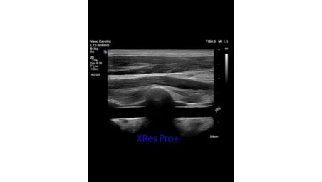
xRes Pro+ uses next-generation image processing to enhance image sharpness and detail resolution, helping provide clear visualization of anatomical structures across a wide range of exams. Advanced processing improves border delineation, particularly in breast, thyroid, and musculoskeletal (MSK) imaging, supporting the assessment of fine anatomical detail. The technology intelligently adjusts imaging parameters based on patient characteristics and penetration requirements to help optimize image quality and consistency across clinical applications.

xRes Pro+

xRes Pro+ video

xRes Pro+ uses next-generation image processing to enhance image sharpness and detail resolution, helping provide clear visualization of anatomical structures across a wide range of exams. Advanced processing improves border delineation, particularly in breast, thyroid, and musculoskeletal (MSK) imaging, supporting the assessment of fine anatomical detail. The technology intelligently adjusts imaging parameters based on patient characteristics and penetration requirements to help optimize image quality and consistency across clinical applications.

xRes Pro+

xRes Pro+ uses next-generation image processing to enhance image sharpness and detail resolution, helping provide clear visualization of anatomical structures across a wide range of exams. Advanced processing improves border delineation, particularly in breast, thyroid, and musculoskeletal (MSK) imaging, supporting the assessment of fine anatomical detail. The technology intelligently adjusts imaging parameters based on patient characteristics and penetration requirements to help optimize image quality and consistency across clinical applications.
Click here for more information
xRes Pro+
xRes Pro+ video

xRes Pro+

xRes Pro+ uses next-generation image processing to enhance image sharpness and detail resolution, helping provide clear visualization of anatomical structures across a wide range of exams. Advanced processing improves border delineation, particularly in breast, thyroid, and musculoskeletal (MSK) imaging, supporting the assessment of fine anatomical detail. The technology intelligently adjusts imaging parameters based on patient characteristics and penetration requirements to help optimize image quality and consistency across clinical applications.
Auto Measure Abdomen
Auto Measure Abdomen video

Auto Measure Abdomen

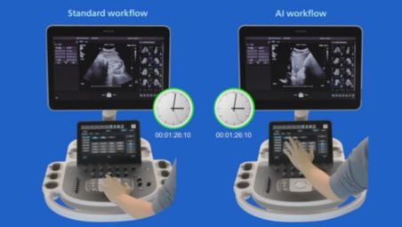
Auto Measure Abdomen is an AI-powered tool that automatically identifies abdominal anatomy and organ borders to place calipers with minimal user input. This helps support more consistent measurements across users while reducing manual steps and streamlining exam workflows.

Auto Measure Abdomen

Auto Measure Abdomen video

Auto Measure Abdomen is an AI-powered tool that automatically identifies abdominal anatomy and organ borders to place calipers with minimal user input. This helps support more consistent measurements across users while reducing manual steps and streamlining exam workflows.

Auto Measure Abdomen

Auto Measure Abdomen is an AI-powered tool that automatically identifies abdominal anatomy and organ borders to place calipers with minimal user input. This helps support more consistent measurements across users while reducing manual steps and streamlining exam workflows.
Click here for more information
Auto Measure Abdomen
Auto Measure Abdomen video

Auto Measure Abdomen

Auto Measure Abdomen is an AI-powered tool that automatically identifies abdominal anatomy and organ borders to place calipers with minimal user input. This helps support more consistent measurements across users while reducing manual steps and streamlining exam workflows.
Philips 2D Auto Cine for effortless frame selection

Philips 2D Auto Cine for effortless frame selection

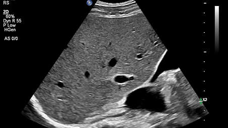
Automatically captures and stores continuous image loops during scanning by recording real-time 2D imaging data. This helps streamline review by allowing users to scroll through captured frames and select relevant moments for documentation, while reducing the need for manual clip acquisition.

Philips 2D Auto Cine for effortless frame selection

Philips 2D Auto Cine for effortless frame selection
Automatically captures and stores continuous image loops during scanning by recording real-time 2D imaging data. This helps streamline review by allowing users to scroll through captured frames and select relevant moments for documentation, while reducing the need for manual clip acquisition.

Philips 2D Auto Cine for effortless frame selection

Automatically captures and stores continuous image loops during scanning by recording real-time 2D imaging data. This helps streamline review by allowing users to scroll through captured frames and select relevant moments for documentation, while reducing the need for manual clip acquisition.
Click here for more information
Philips 2D Auto Cine for effortless frame selection

Philips 2D Auto Cine for effortless frame selection

Automatically captures and stores continuous image loops during scanning by recording real-time 2D imaging data. This helps streamline review by allowing users to scroll through captured frames and select relevant moments for documentation, while reducing the need for manual clip acquisition.
Elastography and Auto ElastQ

Elastography and Auto ElastQ

Philips ultrasound platform supports both strain and shear wave imaging methods of elastography. ElastQ Imaging methods of shear wave elastography use a unique pulsing scheme to generate and detect the propagation speed of shear waves, providing a quantitative display and measurement of tissue stiffness. With Auto ElastQ,experience our next generation of liver health assessment. Auto ElastQ is designed to simplify user workflow with real-time, quantitative shear wave measurements.

Elastography and Auto ElastQ

Elastography and Auto ElastQ
Philips ultrasound platform supports both strain and shear wave imaging methods of elastography. ElastQ Imaging methods of shear wave elastography use a unique pulsing scheme to generate and detect the propagation speed of shear waves, providing a quantitative display and measurement of tissue stiffness. With Auto ElastQ,experience our next generation of liver health assessment. Auto ElastQ is designed to simplify user workflow with real-time, quantitative shear wave measurements.

Elastography and Auto ElastQ

Philips ultrasound platform supports both strain and shear wave imaging methods of elastography. ElastQ Imaging methods of shear wave elastography use a unique pulsing scheme to generate and detect the propagation speed of shear waves, providing a quantitative display and measurement of tissue stiffness. With Auto ElastQ,experience our next generation of liver health assessment. Auto ElastQ is designed to simplify user workflow with real-time, quantitative shear wave measurements.
Click here for more information
Elastography and Auto ElastQ

Elastography and Auto ElastQ

Philips ultrasound platform supports both strain and shear wave imaging methods of elastography. ElastQ Imaging methods of shear wave elastography use a unique pulsing scheme to generate and detect the propagation speed of shear waves, providing a quantitative display and measurement of tissue stiffness. With Auto ElastQ,experience our next generation of liver health assessment. Auto ElastQ is designed to simplify user workflow with real-time, quantitative shear wave measurements.
Quad Imaging
Quad Imaging

Quad Imaging

Quad Imaging supports efficient, consistent AFI assessment by reducing manual steps and helping reduce variability. Designed to support workflow efficiency, it also helps reduce storage requirements across OB practices.

Quad Imaging

Quad Imaging
Quad Imaging supports efficient, consistent AFI assessment by reducing manual steps and helping reduce variability. Designed to support workflow efficiency, it also helps reduce storage requirements across OB practices.

Quad Imaging

Quad Imaging supports efficient, consistent AFI assessment by reducing manual steps and helping reduce variability. Designed to support workflow efficiency, it also helps reduce storage requirements across OB practices.
Click here for more information
Quad Imaging
Quad Imaging

Quad Imaging

Quad Imaging supports efficient, consistent AFI assessment by reducing manual steps and helping reduce variability. Designed to support workflow efficiency, it also helps reduce storage requirements across OB practices.
Koios
Koios

Koios

Koios AI decision support, available with Philips ultrasound both on-cart and off-cart, enables clinicians to classify breast lesions and thyroid nodules. The integration with Koios Bi-RADs offers interpretation and assessment of the risk of malignancy in under 2 seconds, and also leverages Koios Ti-RADS software to support confident lesion classifications using over 350,000 pathology-proven cases.

Koios

Koios
Koios AI decision support, available with Philips ultrasound both on-cart and off-cart, enables clinicians to classify breast lesions and thyroid nodules. The integration with Koios Bi-RADs offers interpretation and assessment of the risk of malignancy in under 2 seconds, and also leverages Koios Ti-RADS software to support confident lesion classifications using over 350,000 pathology-proven cases.

Koios

Koios AI decision support, available with Philips ultrasound both on-cart and off-cart, enables clinicians to classify breast lesions and thyroid nodules. The integration with Koios Bi-RADs offers interpretation and assessment of the risk of malignancy in under 2 seconds, and also leverages Koios Ti-RADS software to support confident lesion classifications using over 350,000 pathology-proven cases.
Click here for more information
Koios
Koios

Koios

Koios AI decision support, available with Philips ultrasound both on-cart and off-cart, enables clinicians to classify breast lesions and thyroid nodules. The integration with Koios Bi-RADs offers interpretation and assessment of the risk of malignancy in under 2 seconds, and also leverages Koios Ti-RADS software to support confident lesion classifications using over 350,000 pathology-proven cases.
Super Resolution MVI with Time of Arrival and Time to Peak
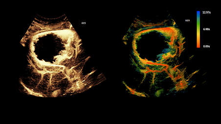

Super Resolution MVI with Time of Arrival and Time to Peak

Enhances visualization of low‑velocity microvascular flow while providing temporal information such as Time of Arrival and Time to Peak. This supports more detailed assessment of flow patterns and helps enable consistent evaluation across users.

Super Resolution MVI with Time of Arrival and Time to Peak

Super Resolution MVI with Time of Arrival and Time to Peak
Enhances visualization of low‑velocity microvascular flow while providing temporal information such as Time of Arrival and Time to Peak. This supports more detailed assessment of flow patterns and helps enable consistent evaluation across users.

Super Resolution MVI with Time of Arrival and Time to Peak

Enhances visualization of low‑velocity microvascular flow while providing temporal information such as Time of Arrival and Time to Peak. This supports more detailed assessment of flow patterns and helps enable consistent evaluation across users.
Click here for more information
Super Resolution MVI with Time of Arrival and Time to Peak

Super Resolution MVI with Time of Arrival and Time to Peak

Enhances visualization of low‑velocity microvascular flow while providing temporal information such as Time of Arrival and Time to Peak. This supports more detailed assessment of flow patterns and helps enable consistent evaluation across users.
Contrast-enhanced ultrasound (CEUS)

Contrast-enhanced ultrasound (CEUS)

CEUS can transform the role of ultrasound in the liver, allowing the study of the enhancement patterns of suspicious liver lesions in real time, as well as providing an alternative non-ionizing approach to the assessment of vesicoureteral reflux in pediatric patients.

Contrast-enhanced ultrasound (CEUS)

Contrast-enhanced ultrasound (CEUS)
CEUS can transform the role of ultrasound in the liver, allowing the study of the enhancement patterns of suspicious liver lesions in real time, as well as providing an alternative non-ionizing approach to the assessment of vesicoureteral reflux in pediatric patients.

Contrast-enhanced ultrasound (CEUS)

CEUS can transform the role of ultrasound in the liver, allowing the study of the enhancement patterns of suspicious liver lesions in real time, as well as providing an alternative non-ionizing approach to the assessment of vesicoureteral reflux in pediatric patients.
Click here for more information
Contrast-enhanced ultrasound (CEUS)

Contrast-enhanced ultrasound (CEUS)

CEUS can transform the role of ultrasound in the liver, allowing the study of the enhancement patterns of suspicious liver lesions in real time, as well as providing an alternative non-ionizing approach to the assessment of vesicoureteral reflux in pediatric patients.
Auto Segmental Wall Motion Scoring

Auto Segmental Wall Motion Scoring

Provides automated evaluation of wall motion in a standard 17-segment bullseye display to aid objective LV wall assessment. With Auto SWMS, you can achieve greater reproducibility and efficiency in your workflows.

Auto Segmental Wall Motion Scoring

Auto Segmental Wall Motion Scoring
Provides automated evaluation of wall motion in a standard 17-segment bullseye display to aid objective LV wall assessment. With Auto SWMS, you can achieve greater reproducibility and efficiency in your workflows.

Auto Segmental Wall Motion Scoring

Provides automated evaluation of wall motion in a standard 17-segment bullseye display to aid objective LV wall assessment. With Auto SWMS, you can achieve greater reproducibility and efficiency in your workflows.
Click here for more information
Auto Segmental Wall Motion Scoring

Auto Segmental Wall Motion Scoring

Provides automated evaluation of wall motion in a standard 17-segment bullseye display to aid objective LV wall assessment. With Auto SWMS, you can achieve greater reproducibility and efficiency in your workflows.
Auto Strain LV with automated EF and mid-layer strain

Auto Strain LV with automated EF and mid-layer strain

Advances to Auto Strain feature fast, reproducible results as part of a comprehensive LV assessment within the same application, improving workflow and saving time. Smart View Select works in the background and uses AI to automatically select the optimum images for 2D LV assessment.

Auto Strain LV with automated EF and mid-layer strain

Auto Strain LV with automated EF and mid-layer strain
Advances to Auto Strain feature fast, reproducible results as part of a comprehensive LV assessment within the same application, improving workflow and saving time. Smart View Select works in the background and uses AI to automatically select the optimum images for 2D LV assessment.

Auto Strain LV with automated EF and mid-layer strain

Advances to Auto Strain feature fast, reproducible results as part of a comprehensive LV assessment within the same application, improving workflow and saving time. Smart View Select works in the background and uses AI to automatically select the optimum images for 2D LV assessment.
Click here for more information
Auto Strain LV with automated EF and mid-layer strain

Auto Strain LV with automated EF and mid-layer strain

Advances to Auto Strain feature fast, reproducible results as part of a comprehensive LV assessment within the same application, improving workflow and saving time. Smart View Select works in the background and uses AI to automatically select the optimum images for 2D LV assessment.
Next Gen Auto Scan
Next Gen Auto Scan

Next Gen Auto Scan

Continuously analyzes image quality in real time and automatically adjusts key imaging parameters during scanning. This helps reduce the need for manual adjustment while also improving transducer plunkability, supporting consistent image acquisition across users and exam types.

Next Gen Auto Scan

Next Gen Auto Scan
Continuously analyzes image quality in real time and automatically adjusts key imaging parameters during scanning. This helps reduce the need for manual adjustment while also improving transducer plunkability, supporting consistent image acquisition across users and exam types.

Next Gen Auto Scan

Continuously analyzes image quality in real time and automatically adjusts key imaging parameters during scanning. This helps reduce the need for manual adjustment while also improving transducer plunkability, supporting consistent image acquisition across users and exam types.
Click here for more information
Next Gen Auto Scan
Next Gen Auto Scan

Next Gen Auto Scan

Continuously analyzes image quality in real time and automatically adjusts key imaging parameters during scanning. This helps reduce the need for manual adjustment while also improving transducer plunkability, supporting consistent image acquisition across users and exam types.
FlexVue with Orthogonal View

FlexVue with Orthogonal View

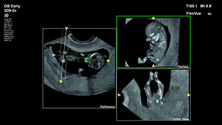
Easy-to-use tools designed to extract challenging anatomical planes from 3D data sets. This advanced feature offers exceptional flexibility in plane acquisition, complemented by a comprehensive measurement package for precise quantification.

FlexVue with Orthogonal View

FlexVue with Orthogonal View
Easy-to-use tools designed to extract challenging anatomical planes from 3D data sets. This advanced feature offers exceptional flexibility in plane acquisition, complemented by a comprehensive measurement package for precise quantification.

FlexVue with Orthogonal View

Easy-to-use tools designed to extract challenging anatomical planes from 3D data sets. This advanced feature offers exceptional flexibility in plane acquisition, complemented by a comprehensive measurement package for precise quantification.
Click here for more information
FlexVue with Orthogonal View

FlexVue with Orthogonal View

Easy-to-use tools designed to extract challenging anatomical planes from 3D data sets. This advanced feature offers exceptional flexibility in plane acquisition, complemented by a comprehensive measurement package for precise quantification.
nSIGHT Imaging
nSIGHT Imaging

nSIGHT Imaging

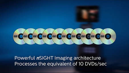
Far surpasses conventional ultrasound performance to reach new levels of definition and clarity. Incorporating a custom multi-stage precision beamformer along with massive parallel processing, this proprietary architecture captures an enormous amount of acoustic data from each transmit operation and performs digital beam reconstruction along with mathematically optimized focal processing. This creates extraordinary real-time images with exceptional frame rate, uniformity and penetration.

nSIGHT Imaging

nSIGHT Imaging
Far surpasses conventional ultrasound performance to reach new levels of definition and clarity. Incorporating a custom multi-stage precision beamformer along with massive parallel processing, this proprietary architecture captures an enormous amount of acoustic data from each transmit operation and performs digital beam reconstruction along with mathematically optimized focal processing. This creates extraordinary real-time images with exceptional frame rate, uniformity and penetration.

nSIGHT Imaging

Far surpasses conventional ultrasound performance to reach new levels of definition and clarity. Incorporating a custom multi-stage precision beamformer along with massive parallel processing, this proprietary architecture captures an enormous amount of acoustic data from each transmit operation and performs digital beam reconstruction along with mathematically optimized focal processing. This creates extraordinary real-time images with exceptional frame rate, uniformity and penetration.
Click here for more information
nSIGHT Imaging
nSIGHT Imaging

nSIGHT Imaging

Far surpasses conventional ultrasound performance to reach new levels of definition and clarity. Incorporating a custom multi-stage precision beamformer along with massive parallel processing, this proprietary architecture captures an enormous amount of acoustic data from each transmit operation and performs digital beam reconstruction along with mathematically optimized focal processing. This creates extraordinary real-time images with exceptional frame rate, uniformity and penetration.
PureWave transducer technology

PureWave transducer technology

Utilizes advanced piezoelectric crystal material to support a wider frequency range, enabling penetration and resolution across a variety of exam types. This helps provide detailed imaging in both deep and superficial structures while supporting consistent image quality.

PureWave transducer technology

PureWave transducer technology
Utilizes advanced piezoelectric crystal material to support a wider frequency range, enabling penetration and resolution across a variety of exam types. This helps provide detailed imaging in both deep and superficial structures while supporting consistent image quality.

PureWave transducer technology

Utilizes advanced piezoelectric crystal material to support a wider frequency range, enabling penetration and resolution across a variety of exam types. This helps provide detailed imaging in both deep and superficial structures while supporting consistent image quality.
Click here for more information
PureWave transducer technology

PureWave transducer technology

Utilizes advanced piezoelectric crystal material to support a wider frequency range, enabling penetration and resolution across a variety of exam types. This helps provide detailed imaging in both deep and superficial structures while supporting consistent image quality.
Auto Doppler
Auto Doppler

Auto Doppler

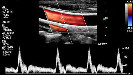
Automatically adjusts color and spectral Doppler settings based on anatomy and flow characteristics, helping reduce manual optimization and support consistent Doppler assessments across users.

Auto Doppler

Auto Doppler
Automatically adjusts color and spectral Doppler settings based on anatomy and flow characteristics, helping reduce manual optimization and support consistent Doppler assessments across users.

Auto Doppler

Automatically adjusts color and spectral Doppler settings based on anatomy and flow characteristics, helping reduce manual optimization and support consistent Doppler assessments across users.
Click here for more information
Auto Doppler
Auto Doppler

Auto Doppler

Automatically adjusts color and spectral Doppler settings based on anatomy and flow characteristics, helping reduce manual optimization and support consistent Doppler assessments across users.
MicroFlow Imaging
MicroFlow Imaging

MicroFlow Imaging

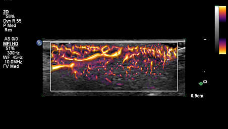
Designed to detect slow and weak blood flow anatomy in tissue. This proprietary approach overcomes many of the barriers associated with conventional methods to detect small vessel blood flow with high resolution and minimal artifacts. MicroFlow Imaging maintains high frame rate and 2D image quality while applying advanced artifact reduction techniques to reveal small vessel anatomy.

MicroFlow Imaging

MicroFlow Imaging
Designed to detect slow and weak blood flow anatomy in tissue. This proprietary approach overcomes many of the barriers associated with conventional methods to detect small vessel blood flow with high resolution and minimal artifacts. MicroFlow Imaging maintains high frame rate and 2D image quality while applying advanced artifact reduction techniques to reveal small vessel anatomy.

MicroFlow Imaging

Designed to detect slow and weak blood flow anatomy in tissue. This proprietary approach overcomes many of the barriers associated with conventional methods to detect small vessel blood flow with high resolution and minimal artifacts. MicroFlow Imaging maintains high frame rate and 2D image quality while applying advanced artifact reduction techniques to reveal small vessel anatomy.
Click here for more information
MicroFlow Imaging
MicroFlow Imaging

MicroFlow Imaging

Designed to detect slow and weak blood flow anatomy in tissue. This proprietary approach overcomes many of the barriers associated with conventional methods to detect small vessel blood flow with high resolution and minimal artifacts. MicroFlow Imaging maintains high frame rate and 2D image quality while applying advanced artifact reduction techniques to reveal small vessel anatomy.
Image Fusion and Navigation-Easy to use modality fusion and interventional guidance
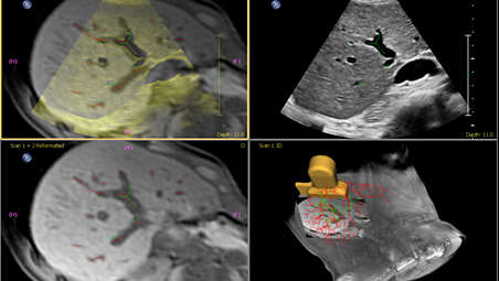

Image Fusion and Navigation-Easy to use modality fusion and interventional guidance

Make confident decisions even in challenging diagnostic cases with fully integrated fusion capabilities that feature streamlined workflows to allow clinicians to achieve fast and effective fusion of CT/MR/PET with live ultrasound. By combining imaging modalities directly on the ultrasound system, you now have access to an even more powerful diagnostic tool with advanced visualization allowing for fast clinical decisions.

Image Fusion and Navigation-Easy to use modality fusion and interventional guidance

Image Fusion and Navigation-Easy to use modality fusion and interventional guidance
Make confident decisions even in challenging diagnostic cases with fully integrated fusion capabilities that feature streamlined workflows to allow clinicians to achieve fast and effective fusion of CT/MR/PET with live ultrasound. By combining imaging modalities directly on the ultrasound system, you now have access to an even more powerful diagnostic tool with advanced visualization allowing for fast clinical decisions.

Image Fusion and Navigation-Easy to use modality fusion and interventional guidance

Make confident decisions even in challenging diagnostic cases with fully integrated fusion capabilities that feature streamlined workflows to allow clinicians to achieve fast and effective fusion of CT/MR/PET with live ultrasound. By combining imaging modalities directly on the ultrasound system, you now have access to an even more powerful diagnostic tool with advanced visualization allowing for fast clinical decisions.
Click here for more information
Image Fusion and Navigation-Easy to use modality fusion and interventional guidance

Image Fusion and Navigation-Easy to use modality fusion and interventional guidance

Make confident decisions even in challenging diagnostic cases with fully integrated fusion capabilities that feature streamlined workflows to allow clinicians to achieve fast and effective fusion of CT/MR/PET with live ultrasound. By combining imaging modalities directly on the ultrasound system, you now have access to an even more powerful diagnostic tool with advanced visualization allowing for fast clinical decisions.
Collaboration Live
Collaboration Live

Collaboration Live

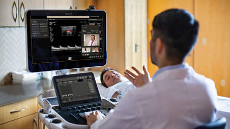
Extend your team without expanding it. Collaboration Live is a communication platform that facilitates communication between a compatible ultrasound system and a remote user. With simultaneous multi-party communication up to six users can quickly and securely talk, text, screen share and video stream directly from the ultrasound system for access to multiple clinical resources at a distance.

Collaboration Live

Collaboration Live
Extend your team without expanding it. Collaboration Live is a communication platform that facilitates communication between a compatible ultrasound system and a remote user. With simultaneous multi-party communication up to six users can quickly and securely talk, text, screen share and video stream directly from the ultrasound system for access to multiple clinical resources at a distance.

Collaboration Live

Extend your team without expanding it. Collaboration Live is a communication platform that facilitates communication between a compatible ultrasound system and a remote user. With simultaneous multi-party communication up to six users can quickly and securely talk, text, screen share and video stream directly from the ultrasound system for access to multiple clinical resources at a distance.
Click here for more information
Collaboration Live
Collaboration Live

Collaboration Live

Extend your team without expanding it. Collaboration Live is a communication platform that facilitates communication between a compatible ultrasound system and a remote user. With simultaneous multi-party communication up to six users can quickly and securely talk, text, screen share and video stream directly from the ultrasound system for access to multiple clinical resources at a distance.
Powerful system security - protecting sensitive patient data
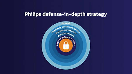

Powerful system security - protecting sensitive patient data

Hospitals and healthcare organizations are spending more to protect their systems and patient data from cyber-attacks. That is why healthcare cybersecurity spending will exceed $65 billion over the next five years. Ultrasound devices are highly mobile and can exist in a wired or wireless environment. As a result, Philips has made security a high priority for ultrasound systems.

Powerful system security - protecting sensitive patient data

Powerful system security - protecting sensitive patient data
Hospitals and healthcare organizations are spending more to protect their systems and patient data from cyber-attacks. That is why healthcare cybersecurity spending will exceed $65 billion over the next five years. Ultrasound devices are highly mobile and can exist in a wired or wireless environment. As a result, Philips has made security a high priority for ultrasound systems.

Powerful system security - protecting sensitive patient data

Hospitals and healthcare organizations are spending more to protect their systems and patient data from cyber-attacks. That is why healthcare cybersecurity spending will exceed $65 billion over the next five years. Ultrasound devices are highly mobile and can exist in a wired or wireless environment. As a result, Philips has made security a high priority for ultrasound systems.
Click here for more information
Powerful system security - protecting sensitive patient data

Powerful system security - protecting sensitive patient data

Hospitals and healthcare organizations are spending more to protect their systems and patient data from cyber-attacks. That is why healthcare cybersecurity spending will exceed $65 billion over the next five years. Ultrasound devices are highly mobile and can exist in a wired or wireless environment. As a result, Philips has made security a high priority for ultrasound systems.
Service
Service

Service

The need to do more with less, rising case complexity and additional care settings put challenges with staffing, skill variability and standardization into sharp focus. This is where our services and solutions can help - get service tailored for your needs with our service contracts, clinical and technical education and training to keep skills fresh, maximize your equipment investment with Technology Maximizer, and extend your team without expanding it with Collaboration Live.

Service

Service
The need to do more with less, rising case complexity and additional care settings put challenges with staffing, skill variability and standardization into sharp focus. This is where our services and solutions can help - get service tailored for your needs with our service contracts, clinical and technical education and training to keep skills fresh, maximize your equipment investment with Technology Maximizer, and extend your team without expanding it with Collaboration Live.

Service

The need to do more with less, rising case complexity and additional care settings put challenges with staffing, skill variability and standardization into sharp focus. This is where our services and solutions can help - get service tailored for your needs with our service contracts, clinical and technical education and training to keep skills fresh, maximize your equipment investment with Technology Maximizer, and extend your team without expanding it with Collaboration Live.
Click here for more information
Service
Service

Service

The need to do more with less, rising case complexity and additional care settings put challenges with staffing, skill variability and standardization into sharp focus. This is where our services and solutions can help - get service tailored for your needs with our service contracts, clinical and technical education and training to keep skills fresh, maximize your equipment investment with Technology Maximizer, and extend your team without expanding it with Collaboration Live.
Education
Education

Education

Our comprehensive education programs are designed to support clinical excellence, increase the use of advanced system features, instill physician confidence in the quality of exams, enhance workflow and productivity, foster professional growth and teamwork, and ultimately deliver an outstanding patient experience.

Education

Education
Our comprehensive education programs are designed to support clinical excellence, increase the use of advanced system features, instill physician confidence in the quality of exams, enhance workflow and productivity, foster professional growth and teamwork, and ultimately deliver an outstanding patient experience.

Education

Our comprehensive education programs are designed to support clinical excellence, increase the use of advanced system features, instill physician confidence in the quality of exams, enhance workflow and productivity, foster professional growth and teamwork, and ultimately deliver an outstanding patient experience.
Click here for more information
Education
Education

Education

Our comprehensive education programs are designed to support clinical excellence, increase the use of advanced system features, instill physician confidence in the quality of exams, enhance workflow and productivity, foster professional growth and teamwork, and ultimately deliver an outstanding patient experience.
  • xRes Pro+
  • Auto Measure Abdomen
  • Philips 2D Auto Cine for effortless frame selection
  • Elastography and Auto ElastQ
See all features
xRes Pro+
xRes Pro+ video

xRes Pro+

xRes Pro+ uses next-generation image processing to enhance image sharpness and detail resolution, helping provide clear visualization of anatomical structures across a wide range of exams. Advanced processing improves border delineation, particularly in breast, thyroid, and musculoskeletal (MSK) imaging, supporting the assessment of fine anatomical detail. The technology intelligently adjusts imaging parameters based on patient characteristics and penetration requirements to help optimize image quality and consistency across clinical applications.

xRes Pro+

xRes Pro+ video

xRes Pro+ uses next-generation image processing to enhance image sharpness and detail resolution, helping provide clear visualization of anatomical structures across a wide range of exams. Advanced processing improves border delineation, particularly in breast, thyroid, and musculoskeletal (MSK) imaging, supporting the assessment of fine anatomical detail. The technology intelligently adjusts imaging parameters based on patient characteristics and penetration requirements to help optimize image quality and consistency across clinical applications.

xRes Pro+

xRes Pro+ uses next-generation image processing to enhance image sharpness and detail resolution, helping provide clear visualization of anatomical structures across a wide range of exams. Advanced processing improves border delineation, particularly in breast, thyroid, and musculoskeletal (MSK) imaging, supporting the assessment of fine anatomical detail. The technology intelligently adjusts imaging parameters based on patient characteristics and penetration requirements to help optimize image quality and consistency across clinical applications.
Click here for more information
xRes Pro+
xRes Pro+ video

xRes Pro+

xRes Pro+ uses next-generation image processing to enhance image sharpness and detail resolution, helping provide clear visualization of anatomical structures across a wide range of exams. Advanced processing improves border delineation, particularly in breast, thyroid, and musculoskeletal (MSK) imaging, supporting the assessment of fine anatomical detail. The technology intelligently adjusts imaging parameters based on patient characteristics and penetration requirements to help optimize image quality and consistency across clinical applications.
Auto Measure Abdomen
Auto Measure Abdomen video

Auto Measure Abdomen

Auto Measure Abdomen is an AI-powered tool that automatically identifies abdominal anatomy and organ borders to place calipers with minimal user input. This helps support more consistent measurements across users while reducing manual steps and streamlining exam workflows.

Auto Measure Abdomen

Auto Measure Abdomen video

Auto Measure Abdomen is an AI-powered tool that automatically identifies abdominal anatomy and organ borders to place calipers with minimal user input. This helps support more consistent measurements across users while reducing manual steps and streamlining exam workflows.

Auto Measure Abdomen

Auto Measure Abdomen is an AI-powered tool that automatically identifies abdominal anatomy and organ borders to place calipers with minimal user input. This helps support more consistent measurements across users while reducing manual steps and streamlining exam workflows.
Click here for more information
Auto Measure Abdomen
Auto Measure Abdomen video

Auto Measure Abdomen

Auto Measure Abdomen is an AI-powered tool that automatically identifies abdominal anatomy and organ borders to place calipers with minimal user input. This helps support more consistent measurements across users while reducing manual steps and streamlining exam workflows.
Philips 2D Auto Cine for effortless frame selection

Philips 2D Auto Cine for effortless frame selection

Automatically captures and stores continuous image loops during scanning by recording real-time 2D imaging data. This helps streamline review by allowing users to scroll through captured frames and select relevant moments for documentation, while reducing the need for manual clip acquisition.

Philips 2D Auto Cine for effortless frame selection

Philips 2D Auto Cine for effortless frame selection
Automatically captures and stores continuous image loops during scanning by recording real-time 2D imaging data. This helps streamline review by allowing users to scroll through captured frames and select relevant moments for documentation, while reducing the need for manual clip acquisition.

Philips 2D Auto Cine for effortless frame selection

Automatically captures and stores continuous image loops during scanning by recording real-time 2D imaging data. This helps streamline review by allowing users to scroll through captured frames and select relevant moments for documentation, while reducing the need for manual clip acquisition.
Click here for more information
Philips 2D Auto Cine for effortless frame selection

Philips 2D Auto Cine for effortless frame selection

Automatically captures and stores continuous image loops during scanning by recording real-time 2D imaging data. This helps streamline review by allowing users to scroll through captured frames and select relevant moments for documentation, while reducing the need for manual clip acquisition.
Elastography and Auto ElastQ

Elastography and Auto ElastQ

Philips ultrasound platform supports both strain and shear wave imaging methods of elastography. ElastQ Imaging methods of shear wave elastography use a unique pulsing scheme to generate and detect the propagation speed of shear waves, providing a quantitative display and measurement of tissue stiffness. With Auto ElastQ,experience our next generation of liver health assessment. Auto ElastQ is designed to simplify user workflow with real-time, quantitative shear wave measurements.

Elastography and Auto ElastQ

Elastography and Auto ElastQ
Philips ultrasound platform supports both strain and shear wave imaging methods of elastography. ElastQ Imaging methods of shear wave elastography use a unique pulsing scheme to generate and detect the propagation speed of shear waves, providing a quantitative display and measurement of tissue stiffness. With Auto ElastQ,experience our next generation of liver health assessment. Auto ElastQ is designed to simplify user workflow with real-time, quantitative shear wave measurements.

Elastography and Auto ElastQ

Philips ultrasound platform supports both strain and shear wave imaging methods of elastography. ElastQ Imaging methods of shear wave elastography use a unique pulsing scheme to generate and detect the propagation speed of shear waves, providing a quantitative display and measurement of tissue stiffness. With Auto ElastQ,experience our next generation of liver health assessment. Auto ElastQ is designed to simplify user workflow with real-time, quantitative shear wave measurements.
Click here for more information
Elastography and Auto ElastQ

Elastography and Auto ElastQ

Philips ultrasound platform supports both strain and shear wave imaging methods of elastography. ElastQ Imaging methods of shear wave elastography use a unique pulsing scheme to generate and detect the propagation speed of shear waves, providing a quantitative display and measurement of tissue stiffness. With Auto ElastQ,experience our next generation of liver health assessment. Auto ElastQ is designed to simplify user workflow with real-time, quantitative shear wave measurements.
Quad Imaging
Quad Imaging

Quad Imaging

Quad Imaging supports efficient, consistent AFI assessment by reducing manual steps and helping reduce variability. Designed to support workflow efficiency, it also helps reduce storage requirements across OB practices.

Quad Imaging

Quad Imaging
Quad Imaging supports efficient, consistent AFI assessment by reducing manual steps and helping reduce variability. Designed to support workflow efficiency, it also helps reduce storage requirements across OB practices.

Quad Imaging

Quad Imaging supports efficient, consistent AFI assessment by reducing manual steps and helping reduce variability. Designed to support workflow efficiency, it also helps reduce storage requirements across OB practices.
Click here for more information
Quad Imaging
Quad Imaging

Quad Imaging

Quad Imaging supports efficient, consistent AFI assessment by reducing manual steps and helping reduce variability. Designed to support workflow efficiency, it also helps reduce storage requirements across OB practices.
Koios
Koios

Koios

Koios AI decision support, available with Philips ultrasound both on-cart and off-cart, enables clinicians to classify breast lesions and thyroid nodules. The integration with Koios Bi-RADs offers interpretation and assessment of the risk of malignancy in under 2 seconds, and also leverages Koios Ti-RADS software to support confident lesion classifications using over 350,000 pathology-proven cases.

Koios

Koios
Koios AI decision support, available with Philips ultrasound both on-cart and off-cart, enables clinicians to classify breast lesions and thyroid nodules. The integration with Koios Bi-RADs offers interpretation and assessment of the risk of malignancy in under 2 seconds, and also leverages Koios Ti-RADS software to support confident lesion classifications using over 350,000 pathology-proven cases.

Koios

Koios AI decision support, available with Philips ultrasound both on-cart and off-cart, enables clinicians to classify breast lesions and thyroid nodules. The integration with Koios Bi-RADs offers interpretation and assessment of the risk of malignancy in under 2 seconds, and also leverages Koios Ti-RADS software to support confident lesion classifications using over 350,000 pathology-proven cases.
Click here for more information
Koios
Koios

Koios

Koios AI decision support, available with Philips ultrasound both on-cart and off-cart, enables clinicians to classify breast lesions and thyroid nodules. The integration with Koios Bi-RADs offers interpretation and assessment of the risk of malignancy in under 2 seconds, and also leverages Koios Ti-RADS software to support confident lesion classifications using over 350,000 pathology-proven cases.
Super Resolution MVI with Time of Arrival and Time to Peak

Super Resolution MVI with Time of Arrival and Time to Peak

Enhances visualization of low‑velocity microvascular flow while providing temporal information such as Time of Arrival and Time to Peak. This supports more detailed assessment of flow patterns and helps enable consistent evaluation across users.

Super Resolution MVI with Time of Arrival and Time to Peak

Super Resolution MVI with Time of Arrival and Time to Peak
Enhances visualization of low‑velocity microvascular flow while providing temporal information such as Time of Arrival and Time to Peak. This supports more detailed assessment of flow patterns and helps enable consistent evaluation across users.

Super Resolution MVI with Time of Arrival and Time to Peak

Enhances visualization of low‑velocity microvascular flow while providing temporal information such as Time of Arrival and Time to Peak. This supports more detailed assessment of flow patterns and helps enable consistent evaluation across users.
Click here for more information
Super Resolution MVI with Time of Arrival and Time to Peak

Super Resolution MVI with Time of Arrival and Time to Peak

Enhances visualization of low‑velocity microvascular flow while providing temporal information such as Time of Arrival and Time to Peak. This supports more detailed assessment of flow patterns and helps enable consistent evaluation across users.
Contrast-enhanced ultrasound (CEUS)

Contrast-enhanced ultrasound (CEUS)

CEUS can transform the role of ultrasound in the liver, allowing the study of the enhancement patterns of suspicious liver lesions in real time, as well as providing an alternative non-ionizing approach to the assessment of vesicoureteral reflux in pediatric patients.

Contrast-enhanced ultrasound (CEUS)

Contrast-enhanced ultrasound (CEUS)
CEUS can transform the role of ultrasound in the liver, allowing the study of the enhancement patterns of suspicious liver lesions in real time, as well as providing an alternative non-ionizing approach to the assessment of vesicoureteral reflux in pediatric patients.

Contrast-enhanced ultrasound (CEUS)

CEUS can transform the role of ultrasound in the liver, allowing the study of the enhancement patterns of suspicious liver lesions in real time, as well as providing an alternative non-ionizing approach to the assessment of vesicoureteral reflux in pediatric patients.
Click here for more information
Contrast-enhanced ultrasound (CEUS)

Contrast-enhanced ultrasound (CEUS)

CEUS can transform the role of ultrasound in the liver, allowing the study of the enhancement patterns of suspicious liver lesions in real time, as well as providing an alternative non-ionizing approach to the assessment of vesicoureteral reflux in pediatric patients.
Auto Segmental Wall Motion Scoring

Auto Segmental Wall Motion Scoring

Provides automated evaluation of wall motion in a standard 17-segment bullseye display to aid objective LV wall assessment. With Auto SWMS, you can achieve greater reproducibility and efficiency in your workflows.

Auto Segmental Wall Motion Scoring

Auto Segmental Wall Motion Scoring
Provides automated evaluation of wall motion in a standard 17-segment bullseye display to aid objective LV wall assessment. With Auto SWMS, you can achieve greater reproducibility and efficiency in your workflows.

Auto Segmental Wall Motion Scoring

Provides automated evaluation of wall motion in a standard 17-segment bullseye display to aid objective LV wall assessment. With Auto SWMS, you can achieve greater reproducibility and efficiency in your workflows.
Click here for more information
Auto Segmental Wall Motion Scoring

Auto Segmental Wall Motion Scoring

Provides automated evaluation of wall motion in a standard 17-segment bullseye display to aid objective LV wall assessment. With Auto SWMS, you can achieve greater reproducibility and efficiency in your workflows.
Auto Strain LV with automated EF and mid-layer strain

Auto Strain LV with automated EF and mid-layer strain

Advances to Auto Strain feature fast, reproducible results as part of a comprehensive LV assessment within the same application, improving workflow and saving time. Smart View Select works in the background and uses AI to automatically select the optimum images for 2D LV assessment.

Auto Strain LV with automated EF and mid-layer strain

Auto Strain LV with automated EF and mid-layer strain
Advances to Auto Strain feature fast, reproducible results as part of a comprehensive LV assessment within the same application, improving workflow and saving time. Smart View Select works in the background and uses AI to automatically select the optimum images for 2D LV assessment.

Auto Strain LV with automated EF and mid-layer strain

Advances to Auto Strain feature fast, reproducible results as part of a comprehensive LV assessment within the same application, improving workflow and saving time. Smart View Select works in the background and uses AI to automatically select the optimum images for 2D LV assessment.
Click here for more information
Auto Strain LV with automated EF and mid-layer strain

Auto Strain LV with automated EF and mid-layer strain

Advances to Auto Strain feature fast, reproducible results as part of a comprehensive LV assessment within the same application, improving workflow and saving time. Smart View Select works in the background and uses AI to automatically select the optimum images for 2D LV assessment.
Next Gen Auto Scan
Next Gen Auto Scan

Next Gen Auto Scan

Continuously analyzes image quality in real time and automatically adjusts key imaging parameters during scanning. This helps reduce the need for manual adjustment while also improving transducer plunkability, supporting consistent image acquisition across users and exam types.

Next Gen Auto Scan

Next Gen Auto Scan
Continuously analyzes image quality in real time and automatically adjusts key imaging parameters during scanning. This helps reduce the need for manual adjustment while also improving transducer plunkability, supporting consistent image acquisition across users and exam types.

Next Gen Auto Scan

Continuously analyzes image quality in real time and automatically adjusts key imaging parameters during scanning. This helps reduce the need for manual adjustment while also improving transducer plunkability, supporting consistent image acquisition across users and exam types.
Click here for more information
Next Gen Auto Scan
Next Gen Auto Scan

Next Gen Auto Scan

Continuously analyzes image quality in real time and automatically adjusts key imaging parameters during scanning. This helps reduce the need for manual adjustment while also improving transducer plunkability, supporting consistent image acquisition across users and exam types.
FlexVue with Orthogonal View

FlexVue with Orthogonal View

Easy-to-use tools designed to extract challenging anatomical planes from 3D data sets. This advanced feature offers exceptional flexibility in plane acquisition, complemented by a comprehensive measurement package for precise quantification.

FlexVue with Orthogonal View

FlexVue with Orthogonal View
Easy-to-use tools designed to extract challenging anatomical planes from 3D data sets. This advanced feature offers exceptional flexibility in plane acquisition, complemented by a comprehensive measurement package for precise quantification.

FlexVue with Orthogonal View

Easy-to-use tools designed to extract challenging anatomical planes from 3D data sets. This advanced feature offers exceptional flexibility in plane acquisition, complemented by a comprehensive measurement package for precise quantification.
Click here for more information
FlexVue with Orthogonal View

FlexVue with Orthogonal View

Easy-to-use tools designed to extract challenging anatomical planes from 3D data sets. This advanced feature offers exceptional flexibility in plane acquisition, complemented by a comprehensive measurement package for precise quantification.
nSIGHT Imaging
nSIGHT Imaging

nSIGHT Imaging

Far surpasses conventional ultrasound performance to reach new levels of definition and clarity. Incorporating a custom multi-stage precision beamformer along with massive parallel processing, this proprietary architecture captures an enormous amount of acoustic data from each transmit operation and performs digital beam reconstruction along with mathematically optimized focal processing. This creates extraordinary real-time images with exceptional frame rate, uniformity and penetration.

nSIGHT Imaging

nSIGHT Imaging
Far surpasses conventional ultrasound performance to reach new levels of definition and clarity. Incorporating a custom multi-stage precision beamformer along with massive parallel processing, this proprietary architecture captures an enormous amount of acoustic data from each transmit operation and performs digital beam reconstruction along with mathematically optimized focal processing. This creates extraordinary real-time images with exceptional frame rate, uniformity and penetration.

nSIGHT Imaging

Far surpasses conventional ultrasound performance to reach new levels of definition and clarity. Incorporating a custom multi-stage precision beamformer along with massive parallel processing, this proprietary architecture captures an enormous amount of acoustic data from each transmit operation and performs digital beam reconstruction along with mathematically optimized focal processing. This creates extraordinary real-time images with exceptional frame rate, uniformity and penetration.
Click here for more information
nSIGHT Imaging
nSIGHT Imaging

nSIGHT Imaging

Far surpasses conventional ultrasound performance to reach new levels of definition and clarity. Incorporating a custom multi-stage precision beamformer along with massive parallel processing, this proprietary architecture captures an enormous amount of acoustic data from each transmit operation and performs digital beam reconstruction along with mathematically optimized focal processing. This creates extraordinary real-time images with exceptional frame rate, uniformity and penetration.
PureWave transducer technology

PureWave transducer technology

Utilizes advanced piezoelectric crystal material to support a wider frequency range, enabling penetration and resolution across a variety of exam types. This helps provide detailed imaging in both deep and superficial structures while supporting consistent image quality.

PureWave transducer technology

PureWave transducer technology
Utilizes advanced piezoelectric crystal material to support a wider frequency range, enabling penetration and resolution across a variety of exam types. This helps provide detailed imaging in both deep and superficial structures while supporting consistent image quality.

PureWave transducer technology

Utilizes advanced piezoelectric crystal material to support a wider frequency range, enabling penetration and resolution across a variety of exam types. This helps provide detailed imaging in both deep and superficial structures while supporting consistent image quality.
Click here for more information
PureWave transducer technology

PureWave transducer technology

Utilizes advanced piezoelectric crystal material to support a wider frequency range, enabling penetration and resolution across a variety of exam types. This helps provide detailed imaging in both deep and superficial structures while supporting consistent image quality.
Auto Doppler
Auto Doppler

Auto Doppler

Automatically adjusts color and spectral Doppler settings based on anatomy and flow characteristics, helping reduce manual optimization and support consistent Doppler assessments across users.

Auto Doppler

Auto Doppler
Automatically adjusts color and spectral Doppler settings based on anatomy and flow characteristics, helping reduce manual optimization and support consistent Doppler assessments across users.

Auto Doppler

Automatically adjusts color and spectral Doppler settings based on anatomy and flow characteristics, helping reduce manual optimization and support consistent Doppler assessments across users.
Click here for more information
Auto Doppler
Auto Doppler

Auto Doppler

Automatically adjusts color and spectral Doppler settings based on anatomy and flow characteristics, helping reduce manual optimization and support consistent Doppler assessments across users.
MicroFlow Imaging
MicroFlow Imaging

MicroFlow Imaging

Designed to detect slow and weak blood flow anatomy in tissue. This proprietary approach overcomes many of the barriers associated with conventional methods to detect small vessel blood flow with high resolution and minimal artifacts. MicroFlow Imaging maintains high frame rate and 2D image quality while applying advanced artifact reduction techniques to reveal small vessel anatomy.

MicroFlow Imaging

MicroFlow Imaging
Designed to detect slow and weak blood flow anatomy in tissue. This proprietary approach overcomes many of the barriers associated with conventional methods to detect small vessel blood flow with high resolution and minimal artifacts. MicroFlow Imaging maintains high frame rate and 2D image quality while applying advanced artifact reduction techniques to reveal small vessel anatomy.

MicroFlow Imaging

Designed to detect slow and weak blood flow anatomy in tissue. This proprietary approach overcomes many of the barriers associated with conventional methods to detect small vessel blood flow with high resolution and minimal artifacts. MicroFlow Imaging maintains high frame rate and 2D image quality while applying advanced artifact reduction techniques to reveal small vessel anatomy.
Click here for more information
MicroFlow Imaging
MicroFlow Imaging

MicroFlow Imaging

Designed to detect slow and weak blood flow anatomy in tissue. This proprietary approach overcomes many of the barriers associated with conventional methods to detect small vessel blood flow with high resolution and minimal artifacts. MicroFlow Imaging maintains high frame rate and 2D image quality while applying advanced artifact reduction techniques to reveal small vessel anatomy.
Image Fusion and Navigation-Easy to use modality fusion and interventional guidance

Image Fusion and Navigation-Easy to use modality fusion and interventional guidance

Make confident decisions even in challenging diagnostic cases with fully integrated fusion capabilities that feature streamlined workflows to allow clinicians to achieve fast and effective fusion of CT/MR/PET with live ultrasound. By combining imaging modalities directly on the ultrasound system, you now have access to an even more powerful diagnostic tool with advanced visualization allowing for fast clinical decisions.

Image Fusion and Navigation-Easy to use modality fusion and interventional guidance

Image Fusion and Navigation-Easy to use modality fusion and interventional guidance
Make confident decisions even in challenging diagnostic cases with fully integrated fusion capabilities that feature streamlined workflows to allow clinicians to achieve fast and effective fusion of CT/MR/PET with live ultrasound. By combining imaging modalities directly on the ultrasound system, you now have access to an even more powerful diagnostic tool with advanced visualization allowing for fast clinical decisions.

Image Fusion and Navigation-Easy to use modality fusion and interventional guidance

Make confident decisions even in challenging diagnostic cases with fully integrated fusion capabilities that feature streamlined workflows to allow clinicians to achieve fast and effective fusion of CT/MR/PET with live ultrasound. By combining imaging modalities directly on the ultrasound system, you now have access to an even more powerful diagnostic tool with advanced visualization allowing for fast clinical decisions.
Click here for more information
Image Fusion and Navigation-Easy to use modality fusion and interventional guidance

Image Fusion and Navigation-Easy to use modality fusion and interventional guidance

Make confident decisions even in challenging diagnostic cases with fully integrated fusion capabilities that feature streamlined workflows to allow clinicians to achieve fast and effective fusion of CT/MR/PET with live ultrasound. By combining imaging modalities directly on the ultrasound system, you now have access to an even more powerful diagnostic tool with advanced visualization allowing for fast clinical decisions.
Collaboration Live
Collaboration Live

Collaboration Live

Extend your team without expanding it. Collaboration Live is a communication platform that facilitates communication between a compatible ultrasound system and a remote user. With simultaneous multi-party communication up to six users can quickly and securely talk, text, screen share and video stream directly from the ultrasound system for access to multiple clinical resources at a distance.

Collaboration Live

Collaboration Live
Extend your team without expanding it. Collaboration Live is a communication platform that facilitates communication between a compatible ultrasound system and a remote user. With simultaneous multi-party communication up to six users can quickly and securely talk, text, screen share and video stream directly from the ultrasound system for access to multiple clinical resources at a distance.

Collaboration Live

Extend your team without expanding it. Collaboration Live is a communication platform that facilitates communication between a compatible ultrasound system and a remote user. With simultaneous multi-party communication up to six users can quickly and securely talk, text, screen share and video stream directly from the ultrasound system for access to multiple clinical resources at a distance.
Click here for more information
Collaboration Live
Collaboration Live

Collaboration Live

Extend your team without expanding it. Collaboration Live is a communication platform that facilitates communication between a compatible ultrasound system and a remote user. With simultaneous multi-party communication up to six users can quickly and securely talk, text, screen share and video stream directly from the ultrasound system for access to multiple clinical resources at a distance.
Powerful system security - protecting sensitive patient data

Powerful system security - protecting sensitive patient data

Hospitals and healthcare organizations are spending more to protect their systems and patient data from cyber-attacks. That is why healthcare cybersecurity spending will exceed $65 billion over the next five years. Ultrasound devices are highly mobile and can exist in a wired or wireless environment. As a result, Philips has made security a high priority for ultrasound systems.

Powerful system security - protecting sensitive patient data

Powerful system security - protecting sensitive patient data
Hospitals and healthcare organizations are spending more to protect their systems and patient data from cyber-attacks. That is why healthcare cybersecurity spending will exceed $65 billion over the next five years. Ultrasound devices are highly mobile and can exist in a wired or wireless environment. As a result, Philips has made security a high priority for ultrasound systems.

Powerful system security - protecting sensitive patient data

Hospitals and healthcare organizations are spending more to protect their systems and patient data from cyber-attacks. That is why healthcare cybersecurity spending will exceed $65 billion over the next five years. Ultrasound devices are highly mobile and can exist in a wired or wireless environment. As a result, Philips has made security a high priority for ultrasound systems.
Click here for more information
Powerful system security - protecting sensitive patient data

Powerful system security - protecting sensitive patient data

Hospitals and healthcare organizations are spending more to protect their systems and patient data from cyber-attacks. That is why healthcare cybersecurity spending will exceed $65 billion over the next five years. Ultrasound devices are highly mobile and can exist in a wired or wireless environment. As a result, Philips has made security a high priority for ultrasound systems.
Service
Service

Service

The need to do more with less, rising case complexity and additional care settings put challenges with staffing, skill variability and standardization into sharp focus. This is where our services and solutions can help - get service tailored for your needs with our service contracts, clinical and technical education and training to keep skills fresh, maximize your equipment investment with Technology Maximizer, and extend your team without expanding it with Collaboration Live.

Service

Service
The need to do more with less, rising case complexity and additional care settings put challenges with staffing, skill variability and standardization into sharp focus. This is where our services and solutions can help - get service tailored for your needs with our service contracts, clinical and technical education and training to keep skills fresh, maximize your equipment investment with Technology Maximizer, and extend your team without expanding it with Collaboration Live.

Service

The need to do more with less, rising case complexity and additional care settings put challenges with staffing, skill variability and standardization into sharp focus. This is where our services and solutions can help - get service tailored for your needs with our service contracts, clinical and technical education and training to keep skills fresh, maximize your equipment investment with Technology Maximizer, and extend your team without expanding it with Collaboration Live.
Click here for more information
Service
Service

Service

The need to do more with less, rising case complexity and additional care settings put challenges with staffing, skill variability and standardization into sharp focus. This is where our services and solutions can help - get service tailored for your needs with our service contracts, clinical and technical education and training to keep skills fresh, maximize your equipment investment with Technology Maximizer, and extend your team without expanding it with Collaboration Live.
Education
Education

Education

Our comprehensive education programs are designed to support clinical excellence, increase the use of advanced system features, instill physician confidence in the quality of exams, enhance workflow and productivity, foster professional growth and teamwork, and ultimately deliver an outstanding patient experience.

Education

Education
Our comprehensive education programs are designed to support clinical excellence, increase the use of advanced system features, instill physician confidence in the quality of exams, enhance workflow and productivity, foster professional growth and teamwork, and ultimately deliver an outstanding patient experience.

Education

Our comprehensive education programs are designed to support clinical excellence, increase the use of advanced system features, instill physician confidence in the quality of exams, enhance workflow and productivity, foster professional growth and teamwork, and ultimately deliver an outstanding patient experience.
Click here for more information
Education
Education

Education

Our comprehensive education programs are designed to support clinical excellence, increase the use of advanced system features, instill physician confidence in the quality of exams, enhance workflow and productivity, foster professional growth and teamwork, and ultimately deliver an outstanding patient experience.

Specifications

Common Specifications
Width
  • 60.6 cm/ 23.9 in
Height
  • 146-171.5 cm/ 57.5-67.5 in
Depth
  • 109.2 cm/ 43 in
Weight
  • 104.3 kg/ 230 lb without peripheral devices
Common Specifications
Width
  • 60.6 cm/ 23.9 in
Height
  • 146-171.5 cm/ 57.5-67.5 in
See all specifications
Common Specifications
Width
  • 60.6 cm/ 23.9 in
Height
  • 146-171.5 cm/ 57.5-67.5 in
Depth
  • 109.2 cm/ 43 in
Weight
  • 104.3 kg/ 230 lb without peripheral devices
  • †Available in select countries. Please consult your Philips representative for further details.
  • *based on a sample size of 20 users
  • ***Compared to previous capability

You are about to visit a Philips global content page

Continue

You are about to visit a Philips global content page

Continue

Our site can best be viewed with the latest version of Microsoft Edge, Google Chrome or Firefox.